1326
Views & Citations326
Likes & Shares
Expansion
in cleft palate can be done by: Symmetrical widening and differential widening.
The orthodontist in his therapeutic armamentarium has several effective
expansion appliances including the W-arch, Quad helix, Hyrax, etc. In selecting
the appliance of choice, certain prerequisites are required like it must be
closely adapted to the palate, it should not be bulky, it should be easy to
clean and it should act as retainer for long period of a time. After closure of
a cleft lip and palate, the patient often experiences a collapse of the
maxillary fragments, resulting in a poor occlusion and an inability to chew
properly. While early surgical
intervention improves the patient’s quality of life lip repair and Closure of
the palatal cleft also tend to constrict the maxilla and produce anterior cross-bite.
The resulting maxillary deficiency is probably the most common problem observed
in such cases.
Keywords: Expansion, Expansion appliance, Cleft lip and
palate
INTRODUCTION
Angle taught that ideal occlusion requires a
full complement of teeth and that an ideal occlusion provides such functional
efficacy that it will be sufficient to ensure a permanent result. Thus arch
expansion to such a degree as to accommodate a full complement of teeth was thought
to be the only way to ensure treatment stability [1].
A major portion of the treatment rendered in
any orthodontic practice is concerned with lack of space – the transverse and
sagittal crowding of teeth within the alveolus. Orthodontic philosophies over
the years have vacillated between a strict non-extraction approach and an
approach, which requires the extraction of teeth [2].
Since the only undisputable yardstick for
measuring success in orthodontic treatment is the appearance and stability of
the case, a substantial time out of all retention has to be given its due
importance [3].
Basic considerations
in maxillary translation
The maxilla can be considered as a bone held
within the facial area of the skull by a series of calcified bony inter
digitations [4], some of which are permanently connected to other bones of the
cranium, such as the frontal, zygomatic, and palatal bones, by connective
tissue fibers which form the suture system. In simple terms the bones can be
moved in any of three ways- laterally, anteriorly or posteriorly but not
medially without extensive resorption of sutural bone margins. Appreciable
lateral movement of the maxilla requires, first, forces sufficiently high to
either exceed the tensile strength of or induced changes in the sutural
connective tissues so that they no longer resist movement of bones, and, second
forces which overcome the extramaxillary muscular and interdigital occlusal
forces. The latter may with the changing relation of bones, then become a
factor contributing to translation of the maxilla.
If the maxilla is to be moved anteriorly, the
same sets of potential resistances are present, but here the interdigitations
of the sutures may come into closer contact so that they oppose movement. In
this situation there are two alternatives; (1) continued application of force
with the objective of inducing resorption of bony serrations so that the two
bones eventually slide across one another; and (2) separation of the bones so
that the sutures no longer interdigitate followed by anterior translation of
the maxilla. The second approach appears to be the logical choice.
The nature of
sutures
Sutures [3-8] are structures joining two
bones by a connective tissue complex which has its peripheral fibers inserted
into the calcified bone margin. Different forms of sutures, adapted to the
local tensions and pressures exerted on bones are found in the skull. Sutures
permit translation of bones and marginal addition of bone tissue during growth
and development as well as movement of bones relative to one another during
muscular function. That is to say, sutures have two functions: (1) they act as
sites of secondary growth; and (2) they provide a shock-absorbing system which
protects the cranial contents during normal bodily function.
The maxillary sutures differ in different
species, at different ages, and range from essentially two thin flat plates of
bone in the young rat to the highly convoluted and interdigitated system in
man. In the latter form, one projection of the bone margin is located within
the socket provided by two projections from the opposing side and the
arrangement of connective tissue fibers is similar to that seen in the
periodontal space.
Changes are produced primarily in the
underlying skeletal structure rather than by the movements of teeth through
alveolar bone. It not only separates the mid palatal suture but also affects
the circumzygomatic and circummaxillary suture system. After the palate is
widened, new bone is deposited in the area of expansion so that the integrity
of the mid palatal suture usually is re- established. Rapid Palatal expansion
is the best example of orthopaedic expansion.
Sutural growth and
its regulation
The majority of the facial and cranial bones
of the vertebrate skull are of intramembranous origin. Growth [5] of these
bones takes place by opposition and resorption at the periosteal surfaces and
by sutural growth. Reshaping of facial and cranial bones occurs by opposition
and resorption at the periosteal surfaces an increase in thickness occurs by a
higher opposition than resorption rate.
Expansion of the skull in a growing
vertebrate is possible by the presence of cranial and facial sutures. The
sutures in the skull have several functions. They unite bones, they absorb
forces, they act as joints that permit relative movement between bones, and
they play a role as growth sites in the growing skull. Troitzky attributed even
bone growth limiting properties to the sutures.
It has been questioned for a long time
whether sutures are autonomous or active growth centers, or whether sutural
growth is adaptive to the growth of surrounding structures. Advocates of the
former view are Massler and Schour, Sicher, Baer and Weinmann and Sicher. These
authors claim the sutures to be autonomous growth centers having intrinsic
growth potency like epiphyseal plates. Growth takes place by sutural tissue
proliferation exerting a separating force on the bony edges. Prahl also
believes that sutures are active growth centers. In an overlapping suture the
fibers connecting the ends of the bony edges run obliquely. Prahl claimed that
shortening of these fibers during maturation can cause separation of the bony
edges of the suture.
The other view is held by such authors as
Gilblin and Alley, Moss, Young, Scott, Moore, Moss and Salentijn and Hoyte.
They consider the suture to be an adaptive growth center, its activity
predominantly determined by surrounding structures.
In the view of Moss, the non-skeletal tissues
and the functioning spaces of the head, like the periosteal matrix and the
growing brain being a capsular matrix, act like functional matrices, which
determine skeletal and consequently sutural growth. Young already explained
this by stating that intracranial pressure forces are converted by the sutural
fibers in tension stimuli, which are known to stimulate osteogenesis. Prahl
however, showed that sutural fibers in the coronal suture of young rats are
directed in such a way that they can with stand extra cranial pressure forces.
Increasing the intracranial pressure initially leads to relaxation of these
fibers. Tensional forces will be exerted on these fibers only when the bony
edges are separated sufficiently.
In the concept of Van Limborgh which is a
synthesis of several earlier theories, sutural growth is controlled by few
intrinsic genetic factors and many local epigenetic factors that originate in
adjacent structures of the head and the skull cartilages. Furthermore, sutural
growth is influenced by local environmental factors occurring in the form of
compressive and tensile forces. General epigenetic and environmental factors
are thought to play only a minor role.
A comparable view is held by Oudhof. The
results of his transplantation experiments suggested that the position and the
structures of sutures are determined hereditarily. Environmental factors
however are required for these characteristics to develop. Furthermore, Enlow
assumed that the genetic information is inadequate to account for the
morphologic complexity found in any given bone. Enlow stated that a suture is a
growth region with its own localized, specialized circumstances, just as all
other parts of the bone have their own regional growth processes.
Response of sutures to extrinsic mechanical forces in vivo
Clinical orthodontic techniques involve the
correction of dentofacial disharmonies by influencing periodontal and sutural
tissues with forces exerted by orthodontic or orthopedic appliances [5].
Sutural tissue response has been studied
mainly in vivo on various types of animals of different ages with use of
different forces and different load/deflection rates. These studies mainly
provide histologic observations of sutural tissue response.
Short-term effects
Investigations of short term effects of force
application can be divided into experiments in which a force is applied
directly to a single suture and experiments in which a force is applied to the
entire maxillary complex.
Direct force
application to a single suture
Ten Cate, Freeman and Dickinson exerted an
expansion force with a spring on the sagittal suture of adult rats. The maximal
opening of the suture was 2 mm. An immediate response consisting of traumatic
tears, exudates, death of fibroblasts, disruption of collagen fibers and acute
inflammation was observed. Although these effects could not always be
demonstrated at the light microscopic level, they were always visible at the
electron microscopic level. Within 3 to 4 days, bone formation was observed at
the edges of the suture, together with collagen deposition and remodeling of
fibroblasts. During diminution and cessation of the expansive force (which took
place within 2 to 3 weeks), remodeling of the bone and the suture occurred
until normal morphology was re-established.
Several
investigators have reported comparable midpalatal sutural responses following
rapid expansion in other animal experiments. Further histologic evidence of
tissue trauma incident to rapid expansion, notably minor fractures of bony
interdigitations, has been presented for monkeys and for human beings.
The severity of
the trauma is related to the increase in sutural width, which in turn is
related to the magnitude of the exerted force. Following force application,
bone formation starts at the bony edges of sutures. The newly formed trabeculae
reflect the direction of the expansive force. Ten Cate, Freeman and Dickinson
noticed that the first bone, which was formed 3 to 4 days after force
application, was laid down in lamellae along the sutural edges. Debbane applied
expansion forces to the palatal suture of full grown casts. Woven bone was
deposited perpendicular to the matrix of existing bone. Deposition in the
suture was unevenly distributed.
Only a few
studies are known in which an attempt was made to quantify the relationship
between external forces and the resulting tissue response in a suture. In most
studies the main objectives were to qualify the ultimate effect of palatal
expansion on the midpalatal suture or to describe the effect of extraoral
forces applied to the maxillofacial complex, including the sutures.
These effects
have been studied in different sutures, in different species, and at different
ages. The observations were predominantly of comparative and descriptive
histologic nature and have dealt mainly with soft rather than bony tissues.
Adaptation of
sutures to altered functional demands can also be observed in transplantation
experiments. These studies showed that although a suture has the capacity to
grow rather autonomously, sutural bone growth is adaptive to environmental
demands. It is understood that an initial traumatic response takes place in the
sutures after application of a force. Some authors suggest that this response
gives relief to internal stresses and strains that are induced by force application.
After the initial response, a period of
growth of the sutural connective tissue takes place. Growth at the bony edges
takes place to reestablish the original sutural morphology. Until now little
has been known about the mechanisms by which forces are transduced into
cellular activity. However, we do know that the response of sutural tissues to
mechanical forces is affected by duration and direction of the force,
morphology of the suture, and age of the subject.
It is still unknown whether all sutures react
in a comparable manner to a given force. In other words, it is still unknown
whether (and if so, to what extent) a close response relationship exists
between applied forces and resulting biologic tissue response in a suture.
An accurate
study of the biologic response of a suture to a force system requires that all
aspects of the force system be identified. In
vivo conditions, it seems impossible to control all force variables that may influence the force system. The
complex morphology of full grown
sutures makes it difficult to predict how a force is dissipated in a suture.
These difficulties in studying the relationship between suture response and
force system variable in vivo suggest
the value of developing an accurate in
vitro model. Because of the reported traumatic responses, it is important
to keep the sutural complex in culture under vital conditions for at least 1
week. With the aid of such an in vitro model, the existence and quality of a
relationship between dose and response might be demonstrated. Information
gathered from experiments in organ
culture under such conditions will lead to a better understanding of sutural
growth and its regulation.
The morphologic and
biochemical effects on tensile force application to the interparietal suture of
the Sprague-Dawley rat
A study was
performed to correlate the histologic and biochemical responses of
interparietial suture to a range of tensile forces [6]. Stainless steel spring
implants, calibrated to generate expensive forces from 50 to 250 g, were placed
across the interparietal suture in 85 female Sprague-Dawley rats. After
experimental periods from 2 h to 14 days, the interparietal sutures were
evaluated by radiography, histology and biochemistry. An in vivo/in vitro system
was used for the biochemical analysis; total protein, proline incorporated,
presence of collagen and alkaline phosphatase activity were measured. The
radiographs and histological evaluation showed that in vivo suture expansion was achievable with 50 to 70 g of force,
but heavier forces showed greater sutural opening, more cellular proliferation,
and more bone formation. This increase in biological response by the heavier
forces was substantiated by an increase in sutural protein and alkaline
phosphatase activity but not in percent collagen. It was concluded that changes
in the total protein content of a suture were not primarily caused by proliferation of osteogenic cells and
fibroblasts but due to an influx of transudate. In contrast, the increase in
incorporation of 3H-proline and alkaline phosphatase activity correlated with
the observance of bone formation. This study indicated a positive correlation
between the magnitude of tensile forces and osteogenic response.
Studies of
orthopedic expansion of cranial sutures have carefully documented that the
biologic response is a widening of the suture followed by the production of
connective tissue components (Hinrichson and Storey, Storey, Cleall and
associates, Murray and Cleall, Ten Cate, Freeman and Dickinson). By this
remodeling activity, the suture reestablishes a configuration similar to its
original form. The remodeling appears to be chemically mediated, but this is
not well understood. Because collagen is the major protein of connective
tissue, the metabolism of this molecule is
important in understanding how tensile forces function.
Cleft is a congenital
anomaly with a multifactorial etiology. The human embryo is most vulnerable
influences during the first trimester of intrauterine life. Any inductive
mechanism, genetic or environmental, that disrupts the organogenetic sequence
in fetal development will cause a congenital malformation.
The cleft
palate patients require extensive and prolonged treatment from birth to
adulthood. This is one area where team approach is very necessary and an
orthodontist plays a major role along with an oral surgeon and prosthodontist.
Cleft palate
patients exhibit a variety of dental irregularities, the severity of which will
be determined by the extent of the deformity and the manner by which it was
surgically repaired.
Maxillary
incisors rotations, collapsed dental arches, congenitally missing teeth,
supernumerary teeth in the cleft area, ectopically erupted teeth, hypoplasia of
labial surfaces of the maxillary incisors, arch length deficiencies,
anteroposterior vertical problems and midline deviation may be found.
Cross-bites in
complete clefts of the lip and palate often have certain characteristics. The
medial displacement of the maxillary segments is usually much more severe in
the canine region and indeed the molars may be in correct lateral relationship.
There is also frequently a deficiency of vertical development, which is worst
in the canine region just behind the cleft. Thus there is often a need to widen
the upper arch considerably in the canine region but hardly at all in the molar
region; in other words to produce differential expansion.
Expansion in
cleft palate can be done by:
1. Symmetrical
widening
2. Differential
widening [7,8]
Symmetrical widening
The
orthodontist in his therapeutic armamentarium has several effective expansion
appliances including the W-arch, Quad helix, Hyrax, Haas expansion screws to
resolve constricted maxillary arches. Of which ‘W’ arch and Quad helix can
cause some amount of differential expansion. In selecting the appliance of
choice, certain prerequisites are required like it must be closely adapted to
the palate, it should not be bulky, it should be easy to clean and it should
act as retainer for long period of a time. The ‘W’ arch and Quad helix seems to
offer most of the requirements and soon also does not affect the speech of the
patients. The only problem may be the amount of expansion possible as some of
the patients have a completely collapsed arch.
Differential
expansion
A differential palatal expansion appliance
was described by Foster and Chinn [9] in 1977, which they modified in 1982. The
appliance consists of a cast metal splint on each segment, joined by an
expansion screw set in a circular housing on each side; the housing in turn is
being fixed to the splints by means of locking plates. The screw is set well forward
between the canine tooth and the circular housing consists of swivel joint
which allows the segments to rotate. A fan type screw can also be used.
The
modification to prevent molar expansion consists
of a palatal bar which is linked to the splints in such a way that it allows the segment to rotate but does not allow
increase of the intermolar width. The palatal bar is formed from a lingual bar,
oval in cross section. Loops of 0.7 mm stainless wire are soldered to the
splints on each side in the molar region and these are linked through 6 gauge
half round clasp wire soldered to the ends of the palatal bar. In case of more
severe discrepancies the screw can be easily changed without much relapse. The
advantages of this appliance over the traditional appliance are:
It is capable of
expanding the canine region more than the molar region
1. It
produces rapid expansion in order to reduce treatment time
2. It is
useful in cases where large range of action is required
3. It
can be kept clean
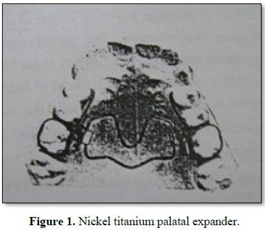
Use of nickel
titanium palatal expander in cleft-palate cases
After closure of a cleft lip and palate, the
patient often experiences a collapse of the maxillary fragments, resulting in a
poor occlusion and an inability to chew properly. While early surgical
intervention improves the patient’s quality of life lip repair and Closure of
the palatal cleft also tend to constrict the maxilla and produce anterior
cross-bite.
The resulting maxillary deficiency is probably
the most common problem observed in such cases (Figure 1).
Although transverse expansion of the maxilla
has been used by orthodontists for more than a century to correct maxillary
anomalies, it can be extremely difficult to use cleft-palate patients.
Many clinicians rely on some form of rapid or
slow palatal expansion for maxillary transverse corrections. Conventional
palatal expanders, besides being uncomfortable for the patients, may require
labor-intensive laboratory construction. Furthermore, the intermittent force
application makes them inefficient, and they are often attached to maxillary
first molars with preexisting mesiolingual rotations that the devices are
unable to correct. Such rotation can distort the appliances, wasting much of
the potential expansion time until the rotations are corrected.
The Nickel palatal Expander [10], developed
by Arndt, produces light, continuous pressure against the midpalatal suture
while simultaneously uprighting, rotating and distalizing the maxillary first
molars. The action of the appliance is a consequence of Nickel titanium’s shape
memory and transition temperature effects. Activated by body temperature, the
Nickel palatal expander automatically expands to its predetermined shape,
requiring little manipulation by the clinician and permitting the patient to
mitigate the pressure, if necessary, by drinking a cold liquid.
As with the NPE, the shape memory of the
Trans palatal wire is activated by temperature. Below the transition point of
94°F, the metal is flexible enough for bending. After insertion, as the
patient’s mouth warms the wire, it tends to return to its original shape. The
light continues force exerted by the wire assures patient comfort. The cleft
palate cases can successfully treated with various sizes and versions of the
Nickel palatal expander, using force levels between 230 g and 300 g. The
appliance also rotates and distalized the maxillary first molars, but no
attempt was made to calculate either the amount of distalization or the
relative amounts of orthopedic versus orthodontic expansion.
SUMMARY AND CONCLUSION
To summarize the different directions and
methods of dental expansion have certain characteristics in common that related
to the technique of expansion and anatomical consideration. All together all
types of maxillary expansion, anterior, lateral and posterior are more
successful easier to perform and less subject to relapse than are the same
procedures in the mandible. Less alveolar bone is present in the anterior
segment of the mandibular dental arch than in the maxillary therefore there is
lesser degree of freedom to move the teeth. In the posterior segments the ramus
reduces the retromolar pad and permits leeway for posterior expansion
1. Paul MD, McNamara JA (1982) Arch width development
in class II patients treated with the Frankel appliances. Am J Orthod 82:
10-12.
2. Haas AJ (1970) Palatal expansion. Just the
beginning of dentofacial orthopedics. Am J Orthod 57: 219-255.
3. Haas AJ (1980) Long term post treatment evaluation
of rapid palatal expansion. Am J Orthod 78: 189-192.
4. Elsdon S (1973) Tissue response to the movement of
bones. Am J Orthod 64: 229-246.
5. Paul AH, Wagemans M (1988) Suture and forces: A
review. Am J Orthod Dentofacial Orthop 94: 29-41.
6. Stanley F, David F (1987) The morphologic and
biochemical effects of tensile force application to the interparietal suture of
the Sprague-Dawley rat. Am J Orthod Dentofacial Orthop 92: 123-133.
7. Proffit WR (1986) Contemporary orthodontics. St
Louis, Mosby Publications, 3rd Edn, 239: 619-621.
8. Chris P (1990) Orthodontic management of the
congenital cleft palate patients. Dent Clin N Am 34: 343-346.
9. Foster JD, Chinn S (1977) Differential rapid
palatal expansion in cleft palate patients. Br J Orthod 143: 139-141.
10. Canoklioghe M (2004) Use of a nickel titanium
palatal expander in cleft palate cases. J Clin Orthod 38: 374-377.
QUICK LINKS
- SUBMIT MANUSCRIPT
- RECOMMEND THE JOURNAL
-
SUBSCRIBE FOR ALERTS
RELATED JOURNALS
- Journal of Infectious Diseases and Research (ISSN: 2688-6537)
- Journal of Neurosurgery Imaging and Techniques (ISSN:2473-1943)
- Journal of Ageing and Restorative Medicine (ISSN:2637-7403)
- Advance Research on Alzheimers and Parkinsons Disease
- Chemotherapy Research Journal (ISSN:2642-0236)
- Journal of Psychiatry and Psychology Research (ISSN:2640-6136)
- Journal of Rheumatology Research (ISSN:2641-6999)
")
")


